Introduction
To see the whole slide imaging (WSI), please register to pathpresenter for once.
There is no charge for this registration.
History
35 yo, F
Nodular lesion on the gingiva between her lower right premolars
Gross Findings
The specimen consisted of a 1.5-cm dome-shaped pale grey soft tissue mass with a smooth, intact surface. It was bisected longitudinally and entirely submitted.
Microscopic Findings
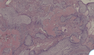
Please click here to see Whole Slide Imaging, then please click the ‘CASE INFO’ button for the explanation
Final Diagnosis:
PERIPHERAL-DENTINOGENIC GHOST CELL TUMOUR
- Quite rare
- The usual presentation is a nodular non-specific swelling
- Share same histopathological features as their intraosseous counterpart
Treatment
- Conservative excision is an appropriate treatment
- Rare recurrence
Take-Home Message
- This lesion seems to be less aggressive than its central counterpart with rare recurrences after simple excision
- Special care must be taken to distinguish dentinogenic ghost cell tumour from ‘ameloblastoma with ghost cells’:
- The proportion of ghost cells (> 1-2%) and the presence of dentinoid are important features in establishing the diagnosis of this entity
