Please click here to see Whole Slide Imaging, then please click the ‘CASE INFO’ button for the explanation
Introduction
To see the whole slide imaging (WSI), please register to pathpresenter for once.
There is no charge for this registration.
History
70 yo, F
Swelling
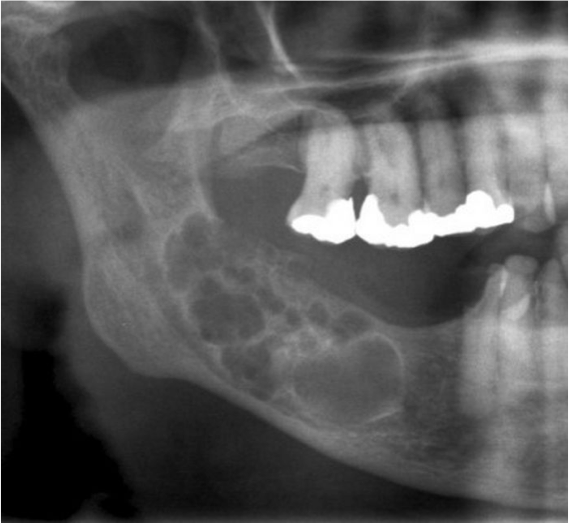
Multilocular radiolucency involving the posterior mandible
Gross Findings
The incisional biopsy consisted of a 1 cm in the area of the lesion. It was bisected longitudinally and entirely submitted.
Microscopic Findings
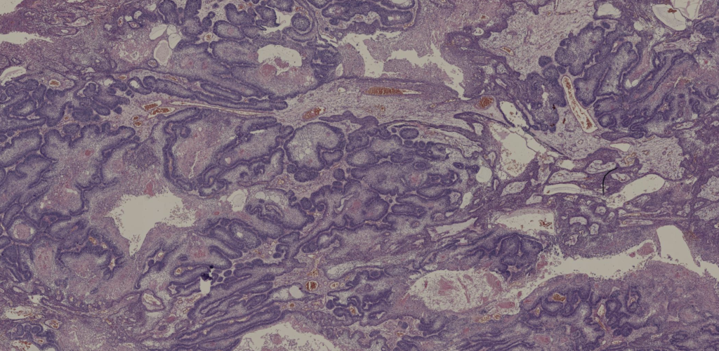
Final Diagnosis:
AMELOBLASTOMA
- The most common odontogenic tumour, excluding odontomas
- age range of 8–92 years and no sex predilection
- Approximately 80%: mandible; posterior region,
- Followed by the anterior mandible, posterior maxilla, and anterior maxilla
- Desmoplastic ameloblastoma: the anterior region of the jaws, especially the maxilla
Treatment
- Wide surgical excision with usually 1.5 cm beyond the radiographic margin
- Conservative surgery: A high recurrence rate
- BRAF inhibitor treatment has been proposed, alone or in combination with MAPK/ERK kinase inhibitors
Take-Home Messages
- Ameloblastoma is a benign but locally infiltrative epithelial odontogenic neoplasm
- No cytological atypia
- It may develop multiple growth patterns (unicystic, multicystic-solid/conventional, peripheral) and histopathological variants (basaloid, acanthomatous, follicular, plexiform, desmoplastic, granular).
- Most tumors have a mixture of histopathological variants.
- The standard of care is complete excision with negative margins irrespective of the histopathological variants.