[vc_row][vc_column icons_position=”left”][rev_slider alias=”cotmoctober”][vc_empty_space height=”2em” alter_height=”none” hide_on_desktop=”” hide_on_notebook=”” hide_on_tablet=”” hide_on_mobile=””][/vc_column][/vc_row][vc_row][vc_column icons_position=”left”][vc_tta_tour style=”modern” active_section=”1″][vc_tta_section title=”Introduction” tab_id=”1609953729115-1d927aec-037efaf2-cdceaf38-ba8b”][vc_column_text]
Introduction
To see the whole slide imaging (WSI), please register to pathpresenter for once.
There is no charge for this registration.
[/vc_column_text][/vc_tta_section][vc_tta_section title=”History” tab_id=”1608318336181-b3708eea-18a6faf2-cdceaf38-ba8b”][vc_column_text]History
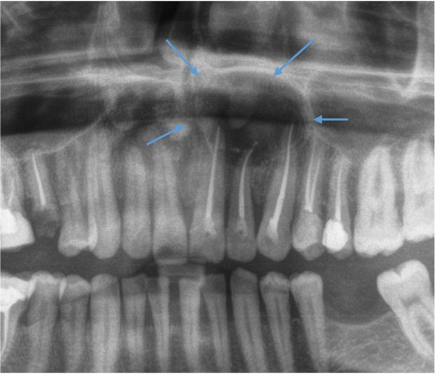
51 yo, M
A well-demarcated, corticated radiolucency at the apex of upper incisor teeth.
[/vc_column_text][vc_single_image image=”1239″ img_size=”full” alignment=”center”][/vc_tta_section][vc_tta_section title=”Gross Findings” tab_id=”1608318336265-4197caba-77f1faf2-cdceaf38-ba8b”][vc_column_text]Gross Findings
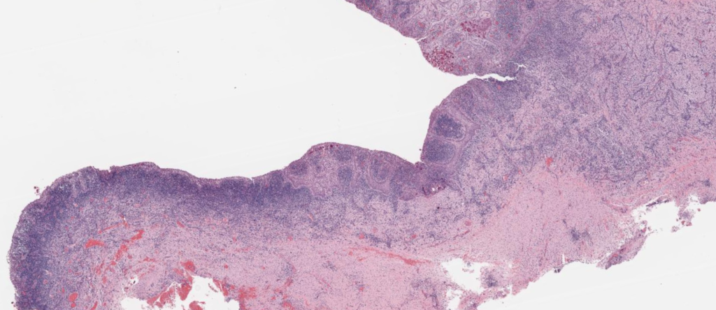
The biopsy consisted of about 1 cm sac-like cystic mass.
[/vc_column_text][/vc_tta_section][vc_tta_section title=”Microscopic Findings” tab_id=”1608318336314-a0c555b4-f7c6faf2-cdceaf38-ba8b”][vc_column_text]Microscopic Findings
[/vc_column_text][vc_single_image image=”1240″ img_size=”full” alignment=”center” onclick=”custom_link” img_link_target=”_blank” link=”https://pathpresenter.net/highyield-viewer/cases/9462a4cd6721dbc09762338b64ab254199d087a64ba21be8422a5166b3fc229cljH+for2W3VMo7%2FfzmGPXw==/c498c8cdcf3a2d029d4d37d7611962fc435be1d042f1232246254c39cb56fcbbLBhCtZdp9SDhfZwzj8wHGg==/b3e8c27dcc634a27c8188337b4c0650355008dbfceb6f9e2ade992233c3a053c2%2FXkdON+6NoLwfZdLICSLg==/3fab438cc64288c6a9ad176d3eb54e98bd87ad60324c7d2f50aa4f4465e6e6d0KP10m7IJAinzRZCzmboxPg==”][vc_column_text]Please click here to see Whole Slide Imaging, then please click the ‘CASE INFO’ button for the explanation
[/vc_column_text][/vc_tta_section][vc_tta_section title=”Final Diagnosis” tab_id=”1608318336420-adcbdc08-6556faf2-cdceaf38-ba8b”][vc_column_text]Final Diagnosis:
RADICULAR CYST
- The most common jaw cyst
- accounting for about 60% of all odontogenic cysts
- A peak incidence in the 4th and 5th decades, a slight male predilection
- The most common site: the anterior maxilla ( 40-50%), followed by the lower molar area
Treatment
- Extraction of the causative tooth or root canal treatment remove the cause
- Enucleation of the cyst is rarely followed by recurrence
Take-Home Messages
- Should be associated with the root of a non-vital tooth (carious, root canal treated, or history of trauma)
- Need clinical-radiological evaluation to exclude the diagnosis of inflamed developmental odontogenic cysts
- Long-standing cysts are less inflamed and have a more regular thin epithelium.