Introduction
To see the whole slide imaging (WSI), please register to pathpresenter for once.
There is no charge for this registration.
History
15 yo, F
Asymptomatic patient
Gross Findings
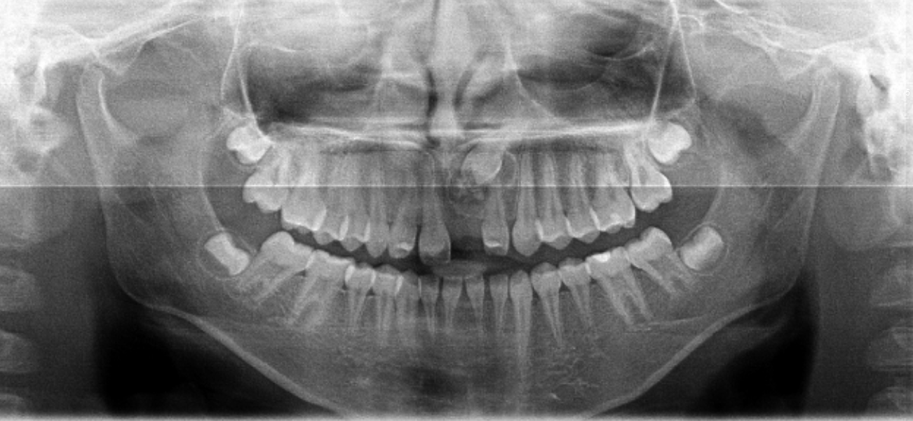
The specimen consisted of an intact central tooth with a cyst-like tissue associated with the crown and half of the root of the tooth.
Microscopic Findings
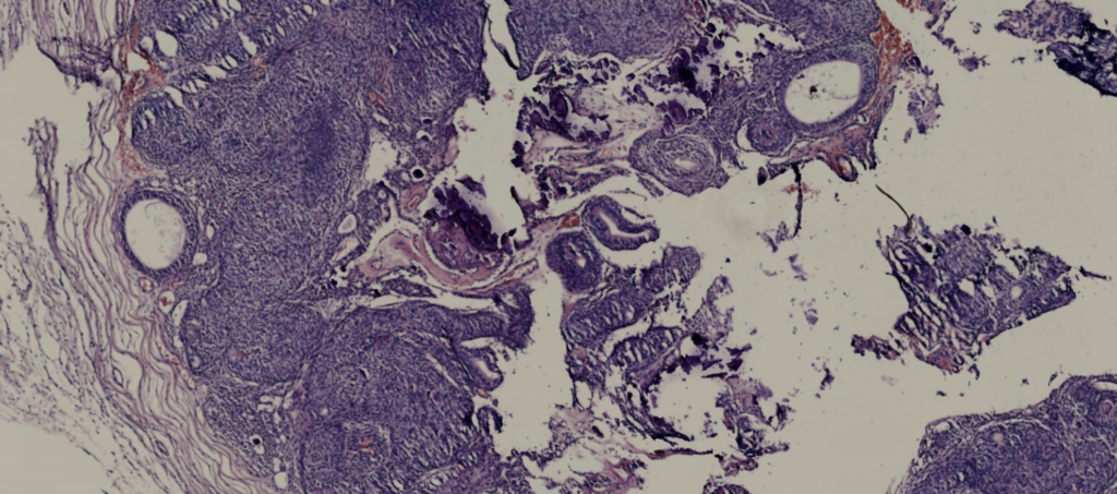
Please click here to see Whole Slide Imaging, then please click the ‘CASE INFO’ button for the explanation
Final Diagnosis:
ADENOMATOID ODONTOGENIC TUMOUR
- < 5% of odontogenic tumours
- More than 95% of AOTs are intraosseous
- Invariably asymptomatic
- Bony expansion +/-
Treatment
- Enucleation
- Recurrence due to incomplete excision
Take-Home Messages
“Two-thirds” rule:
- Two-thirds occur in females
- Two-thirds occur in the maxilla
- Two-thirds are associated with an impacted tooth
- Two-thirds of the impacted teeth are canines
- Two-thirds of the radiographs show radiopaque flecks of calcifications
- Must be differentiated from solid ameloblastoma due to its significantly different biologic behavior and treatment
(Practical Head and Neck Pathology, Frequently Asked Questions, 2019)