Introduction
To see the whole slide imaging (WSI), please register to pathpresenter for once.
There is no charge for this registration.
History
29 yo, F
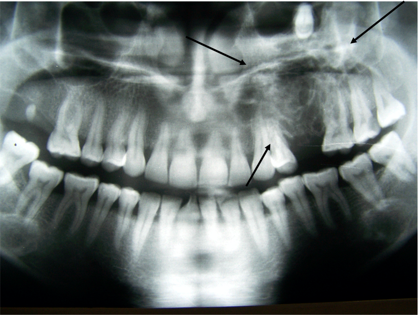
An expansile mixed radiolucent/radiopaque lesion in the posterior maxilla
Gross Findings
The specimen consisted of solid mass fragments.
Microscopic Findings

Please click here to see Whole Slide Imaging, then please click the ‘CASE INFO’ button for the explanation
Final Diagnosis:
CALCIFYING EPITHELIAL ODONTOGENIC TUMOUR
- ≤ 1% of OT
- No gender predilection
- Any age, with a predilection for third to sixth decade of life (mean:40)
- The mandible is affected twice as often as the maxilla
Treatment
- Local surgical removal with tumour-free margin
- Recurrence: 15%
Take-Home Message
- A Congo red stain is useful to confirm the presence of odontogenic ameloblastic associated protein, a substance histologically identical to amyloid
- Due to the rarity of these lesions, pathologists should be aware of the ‘pseudo pleomorphism’ of the tumour cells.
- The Ki-67 index is low in benign lesions, reported as less than 2%. In a case of malignant transformation, the Ki-67 rose to 42% in tumor cells.
