Introduction
To see the whole slide imaging (WSI), please register to pathpresenter for once.
There is no charge for this registration.
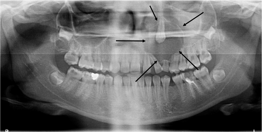
History
21 yo, M
Painless mild swelling
Radiolucent lesion of the anterior maxilla
Gross Findings
The biopsy consisted of multiple strips of cystic epithelium with calcified luminal accretions. Entirely submitted.
Microscopic Findings
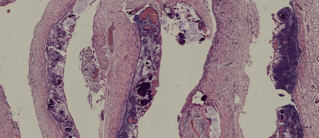
Please click here to see Whole Slide Imaging, then please click the ‘CASE INFO’ button for the explanation
Final Diagnosis:
CALCIFYING ODONTOGENIC CYST
- Rare, accounting for less than 1% of odontogenic cysts
- No gender predilection
- Peak incidence in the second and third decades
- Almost equally in the maxilla and mandible.
- In the maxilla there is a strong predilection for the anterior side
- Approximately 10% of cases are extraosseous with a predilection for the anterior mandibular gingiva
- COC has mutations in CTNNB1, which encodes β catenin
Treatment
- Treated by conservative surgical removal, enucleation and/or curettage.
- 8% recurrence rate
Take-Home Messages
- It exhibits histologic features similar to pilomatrixoma, the epithelium transitions into ghost cell-type keratinization
- Though characteristic, ghost cells form in other odontogenic tumours and do not alone justify a diagnosis of this cyst
- The basal cells resemble ameloblastoma, but there is no microcystic change/cytoplasmic vacuoles between the nuclei and the basement membrane, and the cells are variable cuboidal to columnar, as opposed to tall columnar in ameloblastoma.
- The differential diagnosis includes dentinogenic ghost cell tumor {27669959} and ghost cell odontogenic carcinoma.
