Introduction
To see the whole slide imaging (WSI), please register to pathpresenter for once.
There is no charge for this registration.
History
51 yo, M
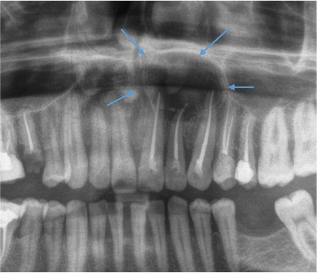
A well-demarcated, corticated radiolucency at the apex of upper incisor teeth.
Gross Findings
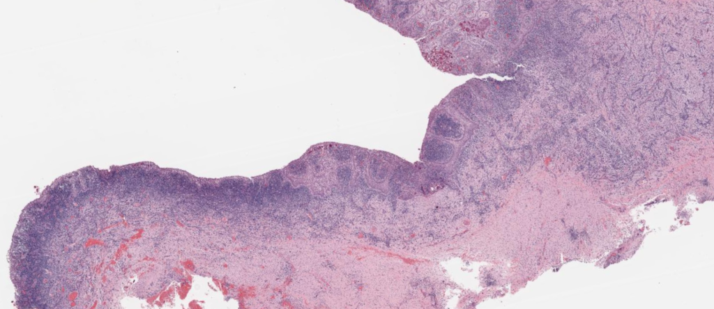
The biopsy consisted of about 1 cm sac-like cystic mass.
Microscopic Findings
Please click here to see Whole Slide Imaging, then please click the ‘CASE INFO’ button for the explanation
Final Diagnosis:
RADICULAR CYST
- The most common jaw cyst
- accounting for about 60% of all odontogenic cysts
- A peak incidence in the 4th and 5th decades, a slight male predilection
- The most common site: the anterior maxilla ( 40-50%), followed by the lower molar area
Treatment
- Extraction of the causative tooth or root canal treatment remove the cause
- Enucleation of the cyst is rarely followed by recurrence
Take-Home Messages
- Should be associated with the root of a non-vital tooth (carious, root canal treated, or history of trauma)
- Need clinical-radiological evaluation to exclude the diagnosis of inflamed developmental odontogenic cysts
- Long-standing cysts are less inflamed and have a more regular thin epithelium.
