Introduction
To see the whole slide imaging (WSI), please register to pathpresenter for once.
There is no charge for this registration.
History
34 yo, M
Incidental radiographic finding
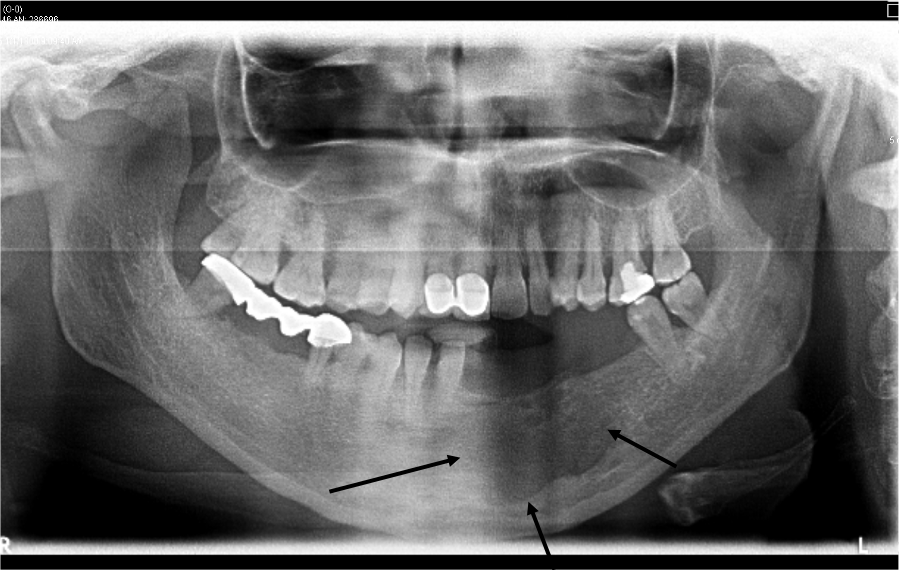
Gross Findings
The specimen consisted of multiple small strips of of sac-like tissue. The specimen was entirely submitted.
Microscopic Findings
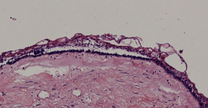
Please click here to see Whole Slide Imaging, then please click the ‘CASE INFO’ button for the explanation
Final Diagnosis:
UNICYSTIC AMELOBLASTOMA
- 5–22% of all ameloblastomas
- Age
- 16 years for cases associated with an impacted tooth
- 35 years in the absence of impaction
- A slight male preponderance
- Most often located in the mandibular third molar area and ascending ramus
- Followed by the body and symphysis
- Most maxillary cases tend to occur in the posterior areas
Take-Home Messages
- Radiographically mimics a cystic lesion,
- Initial treatment often consists of enucleation
- Further treatment is determined by the pattern and extent of the ameloblastomatous proliferation, therefore the whole specimen should be submitted and evaluated microscopically.
2017 WHO :
It has been recommended that a mural type case should be treated as a conventional ameloblastoma when it recurs